
There are several types of medical imaging, but the four most common types are:
X-rays: X-rays use a small amount of ionizing radiation to create images of the body's internal structures. X-rays are commonly used to diagnose bone fractures and to detect the presence of foreign objects in the body.
CT scans: CT scans use a series of X-rays and computer processing to create cross-sectional images of the body. CT scans can provide detailed images of soft tissues, organs, and bones, and are often used to diagnose conditions such as cancer, infections, and injuries.
MRI scans: MRI scans use a powerful magnetic field and radio waves to create detailed images of the body's internal structures. MRI scans are particularly useful for imaging soft tissues such as the brain, spinal cord, and organs, and can be used to diagnose conditions such as tumors, infections, and neurological disorders.
Ultrasound: Ultrasound uses high-frequency sound waves to create images of the body's internal structures. Ultrasound is often used to diagnose conditions such as pregnancy, gallstones, and heart disease.
Each type of medical imaging has its own strengths and limitations, and the choice of imaging modality will depend on the specific needs of the patient and the medical condition being evaluated.
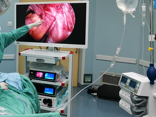
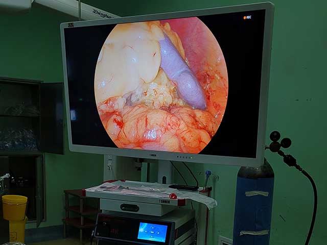
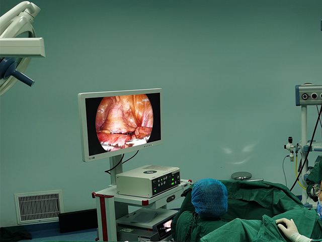
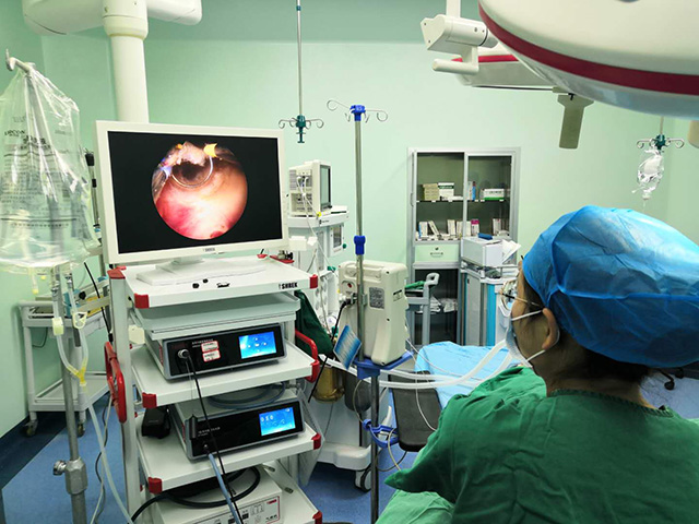
How to present endoscopic surgery images
When presenting endoscopic surgery images, it is important to provide clear and concise information to the audience while ensuring that the images are easily understandable. Here are some tips for presenting endoscopic surgery images:
Provide context: Before showing the images, provide some background information on the patient and the surgical procedure. Explain the purpose of the procedure, the specific structures being examined, and any relevant medical history.
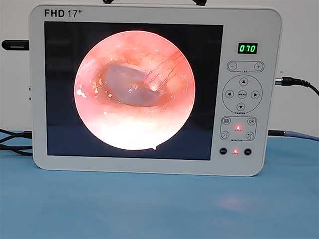
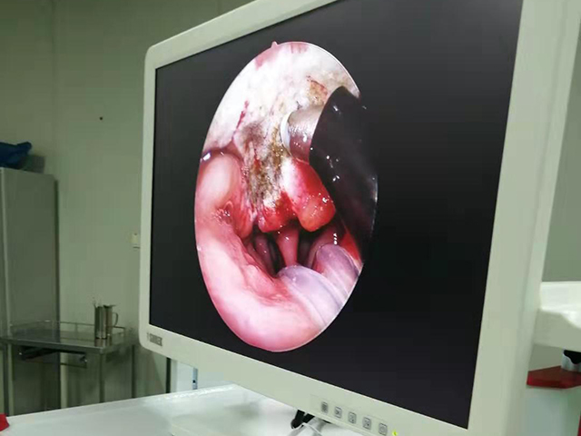
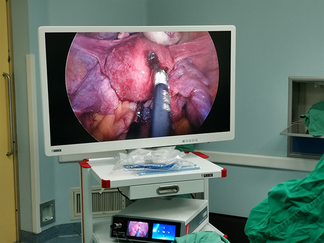
Use clear and high-quality images: Ensure that the images are clear and high-quality so that the audience can easily see the details. If necessary, adjust the lighting or zoom in on specific areas to highlight important features.
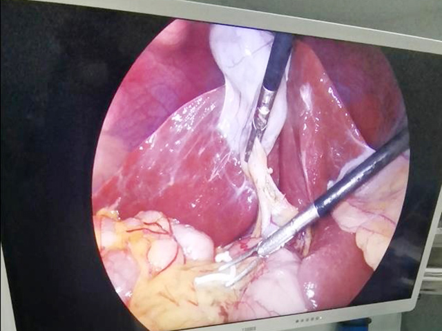
Label and annotate: Label and annotate the images to provide context and clarify what the audience is seeing. Label the specific structures being examined, such as blood vessels, nerves, or organs. Use arrows or circles to highlight specific areas of interest.
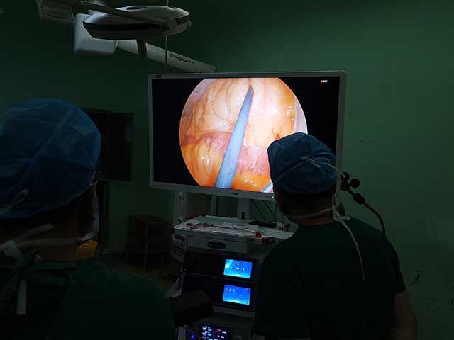
Provide a narrative: Provide a narrative to accompany the images, explaining the steps of the procedure and any findings or observations. This will help the audience understand the significance of the images and the implications for the patient's care.
Use comparison images: If possible, provide comparison images, such as before and after images or images from a healthy patient, to illustrate the changes or improvements resulting from the surgical procedure.
Follow ethical and legal guidelines: Ensure that the presentation follows ethical and legal guidelines regarding patient confidentiality and informed consent. Only use images with the patient's permission and ensure that their identity is protected.
By following these tips, you can effectively present endoscopic surgery images to an audience, whether it is a medical team or a group of students learning about the procedure.

Leave A Inquiry